Main intro Heading link
The Office of the Associate Dean for Research and Graduate Education hosts an annual College of Pharmacy (COP) “Images of Research” competition. The purpose of the competition is to assemble a portfolio of the most innovative and creative images to convey the range of research taking place in COP. These images will be used to promote, advance and represent our College both in printed and digital media.
2023 Image Competition Winners Heading link
148 people voted for the best image in the 13th Annual Images of Research Competition! Congratulations to our winners, whose images are featured below.
View All Original 2023 Images Here
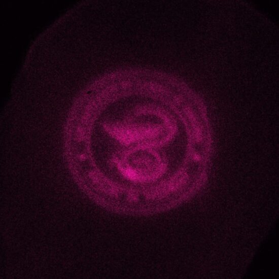
Here we see the College of Pharmacy logo printed on a mouse spleen section with a photoactivatable dye, a digital micromirror device, and blue LED light. I am developing an imaging-based technique for labeling regions of interest to investigate the multiomic changes taking place within the tumor microenviornment and in therapeutic development.
| Department of Pharmaceutical Sciences
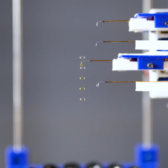
In the Levcell, multiple liquid droplets are suspended in the air using acoustic levitation to counteract the force of gravity. A sample addition arm contactlessly adds reagent (yellow) to previously levitated droplets (clear) in an automated fashion.
| Department of Pharmaceutical Sciences
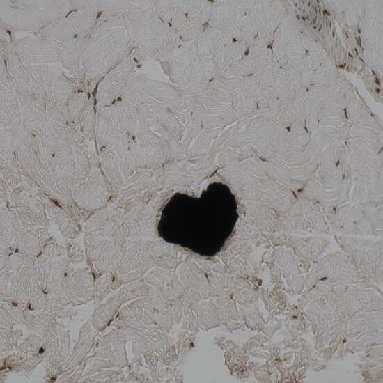
During tissue viability assessment within an ex vivo porcine skin model, the TUNEL assay, designed to identify DNA fragmentation associated with cell apoptosis, indicated that most of the fibroblasts in the lower dermis have undergone apoptosis. But here’s the fun part: we stumbled upon a heart-shaped black spot, probably just some leftover tissue debris, which brought a smile to our faces. It’s a little reminder that science’s surprises can pop up anywhere, even in the most unexpected spots!
| Department of Pharmaceutical Sciences