Image Competition 2015
2015 Image Competition Winners Heading link
Over 300 people voted for the best image in the 5th Annual Images of Research Competition! Congratulations to our winners, whose images are featured below with the number of votes received.
View All Original 2015 Images
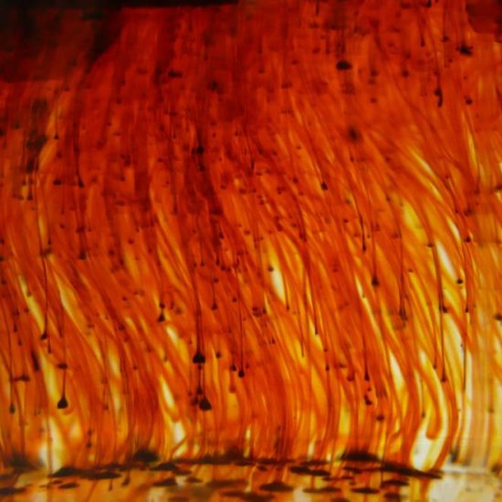
Ordinary moments in research often spawns thought provoking images that capture what is beauty, fear, passion, and awe. Here, malachite green is being used to detect levels of phosphate in enzymatic assays. In making the reagent by adding the salt into sulfuric acid, it flows down as a raw red, reflecting the blood shed from the violence surrounding us in the world. Yet, the distant spaces of white breaking through the image are that small but unyielding image of hope we must actively seek. As researchers, we are reminded that our work is one rooted in our passion to make a difference; one that cannot let negative external forces deter us from moving forward.
| Institute for Tuberculosis Research
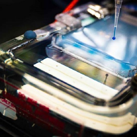
Recently, gene therapy offers a promising therapeutic option for cancer treatment. However, the lack of an efficient gene delivery system largely limits its clinical use. Our goal is to use dendrimer as a carrier to incorporate the gene. Gel electrophoresis is a simple method for the separation and detection of differently sized genes. In our experiment, negatively charged plasmid DNA was incubated with a positively charged dendrimer to prepare a dendrimer-based gene delivery system. Gel electrophoresis was applied to investigate if the gene was firmly packed with dendrimer. The picture was taken at the moment when we were loading samples in the gel electrophoresis apparatus.
| Dept. of Biopharmaceutical Sciences
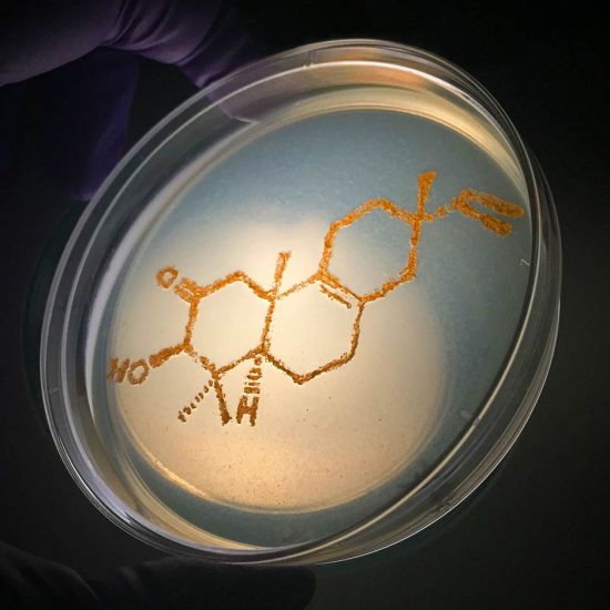
The diterpene class of secondary metabolites is ubiquitous in plants and fungi, but only approximately 20 of these compounds have been reported from bacteria to date. Surprisingly, a novel Δ8,9-pimarane diterpene was isolated in the Murphy lab from an actinomycete bacterium collected in the East Sea of Vietnam. Purified culture of this producing strain was streaked onto a nutrient agar plate in the shape of this molecule’s structure and incubated for seven days, resulting in the orange bond-line drawing shown. The structure elucidation and biological characterization of this molecule and others isolated from this strain were published collaboratively by the Murphy (UIC-COP), Burdette (UIC-COP), and Pham (VAST, Vietnam) labs in the journal ‘Marine Drugs’ in the summer of 2015.
| Dept. of Medicinal Chemistry & Pharmacognosy