Image Competition 2013
2013 Image Competition Winners Heading link
184 people voted for the best image in the 3rd Annual Images of Research Competition! Congratulations to our winners, whose images are featured below with the number of votes received.
View All Original 2013 Images
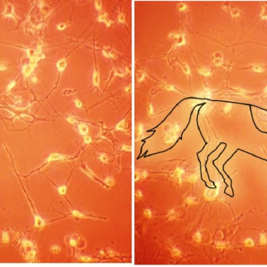
Glioblastoma multiforme is the most common and aggressive form of primary brain tumor in human and accounts for approximately 60% of all diagnosed brain tumors in the United States each year. In our lab, we work with glioblastoma and use hydrogels made up of a variety of polymers as drug delivery vehicles in order to treat this disease. Pictured here are confluent glioblastoma U87-MG cells grown on tissue culture plastic. Can you spot the racing horse outlined by the cells?
| Dept. of Biopharmaceutical Sciences
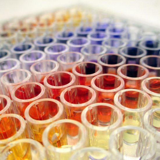
Siderophores are high-affinity iron chelating natural products that are produced by microorganisms. The secretion of siderophores into extracellular environment is crucial for the growth of microorganisms in iron-limiting environments. This picture was captured during the development of a siderophore detection assay. The iron chelating agents under different concentrations rendered a plate of rainbow colors in this assay.
| Dept. of Medicinal Chemistry and Pharmacognos
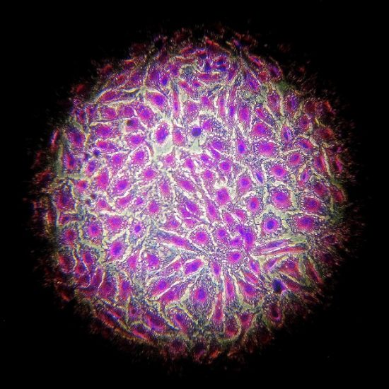
The cells stained with crystal violet are skov-3, a kind of epithelial ovarian cancer cells that derived from human ascites. Originally our aim is to compare the difference in the intensity of the purple color which indicates the growing ability of the cells under different conditions. While observing and magnifying the cells under the microscope, we found the fascinating staining results that can clearly elucidate the structure of the cells.
| Dept. of Biopharmaceutical Sciences