Image Competition 2019
2019 Image Competition Winners Heading link
122 people voted for the best image in the 9th Annual Images of Research Competition! Congratulations to our winners, whose images are featured below with the number of votes received.
View All Original 2019 Images
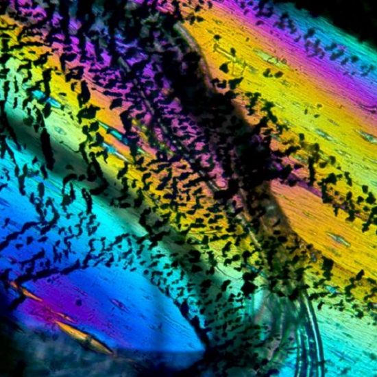
Tapes, like duct tape, are items that most people encounter daily without giving them a second thought. Criminals however, have found nefarious uses for tapes: A tape’s use is limited only by a criminal’s creativity. My research aims to expand upon and optimize the way tapes are analyzed in crime labs through the use a microspectrophotometer. This instrument measures a sample’s electromagnetic absorption in the ultraviolet or visible ranges. More simply, this instrument quantifies color. Before microspectrophotometric observations can be made, an evidentiary tape sample should be analyzed through the crossed polars of a polarized light microscope, providing insight to the tape’s film orientation. The imaged tape, at 100x magnification through cross-polarized light, shows that this duct tape sample was stretched and pulled after manufacture. While this unfortunately meant this sample’s film orientation could not be determined, it did provide this unique and beautiful birefringence pattern.
| Dept. of Pharmaceutical Sciences
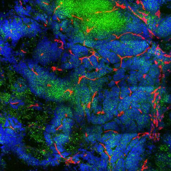
Breast tumor mouse models enable experimental studies which are aided by confocal fluorescence imaging. Here I have used fluorescent antibodies specific to cellular markers to pinpoint tumor cells and surrounding blood vessels. A breast tumor that expresses human epidermal growth factor receptor 2 (Her2) was excised and subjected to an immunostaining and clearing process for whole tumor imaging called Transparent Tissue Tomography (T3). The tumor has been stained with DyLight488-anti-Her2 (green) to identify the membranes of tumor cells, with DyLight633-anti-CD31 (red) to identify blood vessels, and with DAPI (blue) to stain the nuclei of cells. The tumor was made optically transparent by tissue clearing through an ascending gradient of fructose and then imaged with a 40X immersion objective (Zeiss LSM 710). This high-magnification two-dimensional image slice shows where blood vessels are interspersed among Her2-expressing cells. Image analysis of such data is helpful to understand tumor growth and experimental treatment effects.
| Dept. of Pharmaceutical Sciences
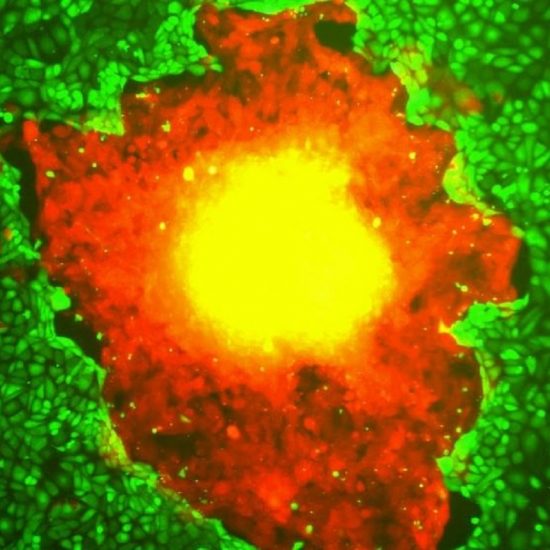
Burdette lab post-doctoral Fellow Dr. Amrita Salvi looks onto mouse derived fallopian tube cells exposed to BPA through a fluorescence microscope. BPA is a synthetic precursor used in the manufacturing of many plastics we use today and has been found to seep into the food and water with which it comes in contact. Recently, BPA has drawn attention as a potential carcinogen through its activity as an endocrine disruptor. The fallopian tube is a known progenitor site of high grade serous ovarian cancer and relies on hormonal signaling to maintain fertility and homeostasis. Pictured, Dr. Salvi is observing fluorescently labeled estrogen receptor (red) localizing to the nucleus (blue) indicating that BPA is exerting aberrant estrogenic activity on the fallopian tube. This finding would suggest a tumorigenic avenue for BPA in the development of ovarian cancer; further research is needed to better understand how BPA may impact women’s reproductive health.
| Dept. of Pharmaceutical Sciences