Image Competition 2012
2012 Image Competition Winners Heading link
Many people voted for the best image in the 2nd Annual Images of Research Competition! Congratulations to our winners, whose images are featured below.
View All Original 2012 Images
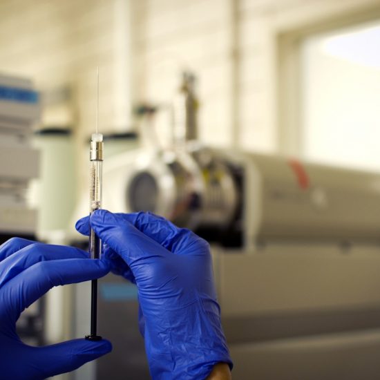
The image captures the process of HPLC/MS run. Pre-clinical pharmacokinetic study of newly discovered compound (to be used in anti-bioterrorism) is done by using HPLC/MS machine. Drug concentration is measured to test its microsomal stability.
| PharmD Student
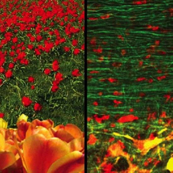
The use of transgenic mice and confocal microscopy methods allow for a detailed study of motor neurons (MNs) in health and disease. This image corresponds to a spinal cord section obtained from a transgenic mouse expressing yellow fluorescent protein (YFP). Stained in red by ethidium bromide, nuclei of oligodendrocytes appear as small roses dispersed amongst a garden of YFP-filled axons pseudocolored in green. At the bottom of this image, large tulip-shaped structures corresponding to cell bodies of MNs are stained orange by the combined colors of YFP and ethidum bromide. Thanks to healthy MNs we can enjoy a walk along a beautiful flower garden!
| Dept. of Medicinal Chemistry & Pharmacognosy
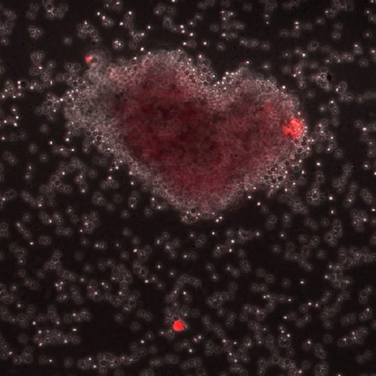
More than a few billion hematological cells, such as leukocytes and red blood cells coexist in one milliliter of human blood. Our objective is to capture rare human disease-related cells among one million-one billion hematological cells. While screening the results, we found a heart-shaped leukocyte aggregation next to the captured target cell (a single red cell). This promising result is not only visible to the eye, but also appeals to the heart.
| Dept. of Biopharmaceutical Sciences