Image Competition 2022
2022 Image Competition Winners Heading link
133 people voted for the best image in the 12th Annual Images of Research Competition! Congratulations to our winners, whose images are featured below with the number of votes received.
View All Original 2022 Images
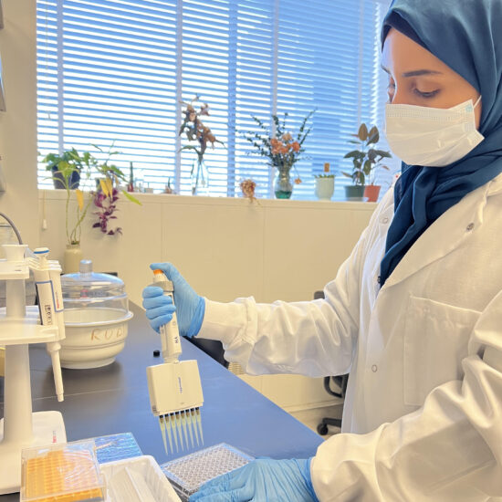
Staining cells for flow cytometry.
| Department of Pharmaceutical Sciences
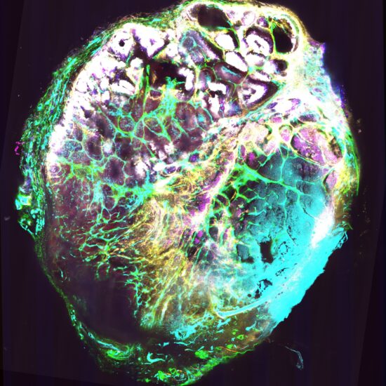
A TUBO tumor slice stained with SMA(red), ER-TR7(green), CD31(cy5), CD8(purple), CD45(yellow),CD3(blue) and CK8(gray). This TUBO tumor was treated with STING agonist by intratumoral injection 48hrs before collection and we applied multiplexed cyclic immunofluorescence for multiple channel images. We can clearly see the TUBO cells were under attack by immune cells.
| Dept. of Pharmaceutical Sciences
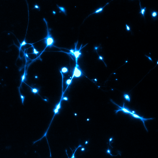
These “Fairy Christmas lights” are produced by the intracellular Fura-2-acetoxymethyl ester (Fura-2 AM) dye in cultured dorsal root ganglion neurons. The Fura-2 AM is a membrane permeable dye, which enters the cells where intracellular esterase activity cleaves the AM esters leaving the dye impermeable and trapped within the cell. Fura-2 is a ratio metric calcium indicator where the excitation spectrum of the calcium sensitive dye is altered upon binding to calcium and can thus indicate an alteration in intracellular calcium. This method allows the visualization of calcium shifts and is used to monitor neuronal activity.
| Dept. of Pharmaceutical Sciences