Image Competition 2020
2020 Image Competition Winners Heading link
119 people voted for the best image in the 10th Annual Images of Research Competition! Congratulations to our winners, whose images are featured below with the number of votes received.
View All Original 2020 Images
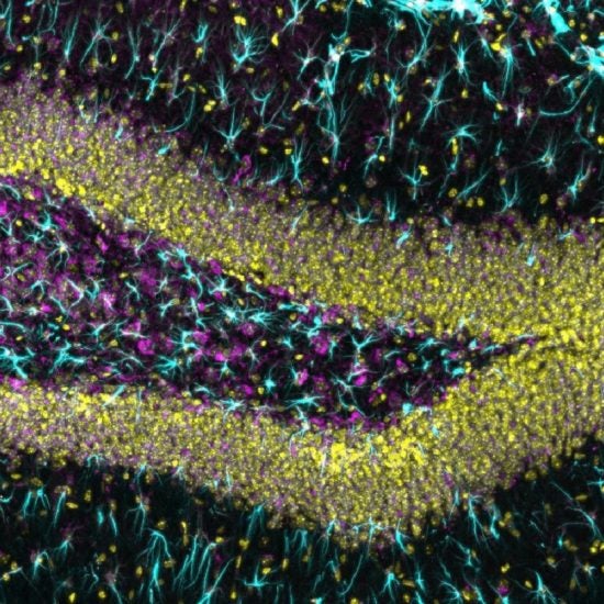
The function of glia cells were underestimated for decades and only in the past 10 years neuroscientists are gaining a deeper understanding of how glia play active roles in modulating neural activity, except for the support function as glia glue. The picture shoot here is the DG region in mouse hippocampus which is responsible for memory storage and potential adult neurogenesis. Cells marked in Cyan are astrocytes which are the most abundant glia type in center nervous system, with Yellow stained in nucleus (DAPI). (Zeiss LSM 710 Confocal, RRC fluorescence Core)
| Dept. of Pharmaceutical Sciences
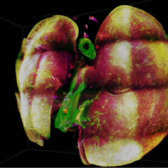
Taking over the duty of oxygen delivery to every single cell in our bodies, lung is the most important part of the respiratory system. Every human being takes more than 6 million breaths per year. However, every breath we take in is accompanied by potentially harmful pathogens that might damage our lungs and cause serious lung disease. In preclinical studies of lung diseases using mouse models, conventional thin tissue imaging assay has drawbacks of lacking 3D information, regional sampling bias, information loss of contiguous structure like airway and vasculature. Therefore, I develop a 3D multiplex fluorescence imaging method for the observation of mouse lung. This method visualizes multiple cell types and biomarkers in a whole mouse lung at single-cell resolution. It maintains the murine lung integrity and visualizes the lung architecture without tissue dissection. Here shows a whole mouse lung image taken by light-sheet microscope with immunofluorescence labels of fibroblasts (green), immune cells (red), and blood vessels (magenta). The 3D microscopy technique will be a powerful imaging tool for preclinical studying of many pulmonary diseases including asthma, lung fibrosis, lung cancer, and COVID-19 infection.
| Dept. of Pharmaceutical Sciences
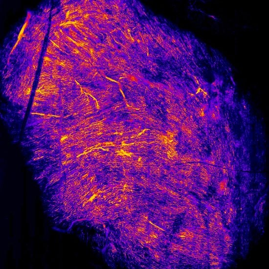
A mouse heart visualized by its “fiery” veins. The coronary veins course along and deep to the surface of the heart. These vessels are essential as they move deoxygenated blood from the working muscle of our heart to the right atrium. From here begins the process to pump the body’s returning deoxygenated blood to the lungs for reoxygenation and removal of carbon dioxide.
| Dept. of Pharmaceutical Sciences